A Fixed-kvp Technique Chart Uses Which of the Following
- Ion chamber - Photocell - Solid state detector. Almost any choice of factors will yield an acceptable image.
Radiographic Exposure Technique Radiology Key
20 mAs at 91 kVp.

. Calculation for determining an approximate amount of kilovoltage kVp necessary for a given anatomic area on the basis of measurement and the grid being used. The kVp is adjusted to compensate. A fixed kVp technique chart D.
Use a fixed mAs variable kVp technique chart. None stated in article. Use a fixed mAs variable kVp technique chart.
What is a major benefit of a fixed kVp technique chart. All factors including kVp are fixed except time. 40 mAs at 91 kVp.
What is a major benefit of a fixed kVp technique chart. Optimum time is found for an average patient. Establishing a fixed kVp technique chart.
Which of the following devices can be used as the sensor in AEC units. Produce initial phantom images - to determine optimal kVp - using abdomen or pelvis phantom. When formulating the technique chart use the same IR grid and processor for each exposure.
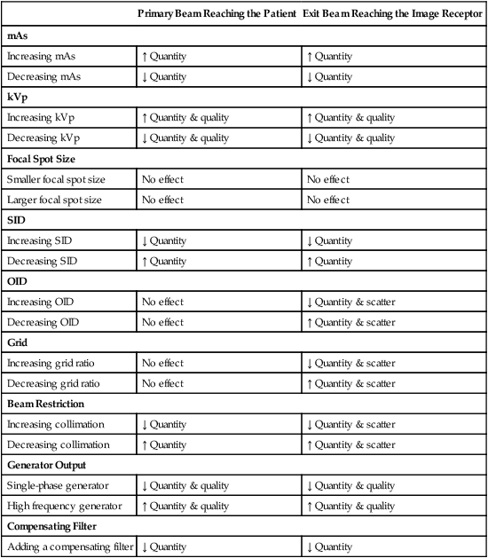
The penetrating quality of the beam for the average adult patient. It is the preferred exposure technique whenever an X-ray machine has independently selectable kVp and mA or mAs settings. Increased tube loading D.
A chart based on tissue thickness and anatomic part that can be consulted for. 80 mAs at 75 kVp. A greater increase is needed for high kVp 90 and above than for low kVp below 70.
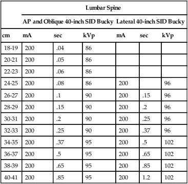
HIPPELVIS Grid mAs CM kVp mAs CMkVp kVp AP HipPelvis Y 15 13-14 72 30 19-20 78 25-26 84 44 225 15-16 72 45 21-22 78 27-28 84 30 17-18 72 60 23-24 78 29-30 84 KNEE APOblq Knee Grid mAs CM kVp Yes 113 7-8 66 150 11-12 70 15-16 70 44 150 9-10 66 225 13-14 7017-18 Lateral Knee Decrease 4 kVp Decrease 4 kVp Decrease 4 kVp LOWER LEG APLateral Grid mAs. 200mA upper limit for small focal spot Thoracic Spine Lumbar Spine Abdomen Skull. A kVp-variable chart uses constant settings for mA and exposure time seconds and.
Getting started You will need to make three test exposures of a medium sized dog. Mammographic image quality contrast and dose for a variable tube potential kVp technique protocol for film-screen mammography have been investigated. A fixed mAs technique chart.
KVp is used to compensate for variations in the thickness of. Select optimal phantom image - based on highest kVp and lowest contrast in acceptance limits process is one of elimination not selecting the best image 3. 2 tissue thickness in cm source-image distance grid factor kVp.
Adequate part penetration B. Identifies reliable exposure factors mA kVp and exposure time for a known tissue thicknesses A radiograph exposure chart can be constructed to suit any X-ray machine. Technique chart construction.
Fixed kVp technique produce long scale contrast and precise part measurement is not required. In this protocol the tube potential is increased for larger breast thicknesses. At the conclusion of this chapter you will be able to.
It requires higher kVp to minimize patient dose. A variable kVp technique chart B. Fixed kVp technique chart uses a fixed optimum kVp and mAs that varies with thickness.
Comparisons were made with fixed kVp protocols in which the tube. What kind of chart uses a kVp value that is high enough to adequately penetrate the part but does not diminish radiographic contrast. 300 mA large focal spot Chest any procedure where.
Fixes kilovoltage technique is aka Optimal or high kVp 6 One a fixed technique chart the kV selection is based on. A and C C. For a thicker than average patient the time is doubled and for a thinner than average patient it is halved.
How much should kVp be increased variable kVp chart for each additional centimeter of tissue to be penetrated. Exposure charts may be kVp-variable or mAs-variable. Proper exposure of a patient to x-radiation is necessary to produce a diagnostic radiograph.
This program uses the kVp-variable method of determining exposures. 40 mAs at 75 kVp. Read and use an x-ray technique chart List methods for obtaining andor creating an x-ray technique chart Accurately measure a body part using an x-ray caliper Compare fixed kilovolts peak kVp technique charts with variable kVp technique charts and state which is preferable.
Decreased exposure latitude C. The objective is that each radiograph should contain an image of the abdomen Figure 1 allowing you to. What technique should be used for the next patient who measures 34cm.
Technical factors must be chosen carefully to yield an acceptable image. Remember that a 15 change in kVp does not produce the same effect across the entire range of kVp used in radiography. A patient measuring 26cm requires 20 mAs at 75 kVp based on a fixed kVp-variable mAs technique chart.
Increase in patient exposure A. In addition whenever a 15 change is made in the kVp to maintain the exposure to the IR the radiographer must adjust the mAs by a factor of 2.

The Settings Of Kvp Mas And Sid In Each Exposure Download Scientific Diagram
The Settings Of Kvp Mas And Sid In Each Exposure Download Scientific Diagram
Formulating X Ray Techniques Radiology Key
No comments for "A Fixed-kvp Technique Chart Uses Which of the Following"
Post a Comment